


Medical imaging is a key part of modern healthcare, helping doctors to diagnose and treat diseases more accurately. Techniques like ultrasonography, X-rays, MRIs, and CT scans let doctors see inside the body to find problems or changes. These tools have changed how doctors understand and treat patients, making medical care better and more precise.
Key Takeaways
- Medical imaging technologies have evolved significantly and continue to advance, offering new ways to diagnose and treat diseases.
- Ultrasonography now includes advanced methods like 3D and 4D imaging, elastography, and specialized uses in cardiology.
- MRI technology has expanded to include functional MRI for brain studies and improved methods for cancer detection.
- CT scans provide high-speed, precise images and are now used for detailed cardiovascular health checks and lung cancer screenings.
- Emerging imaging techniques in cardiovascular diagnostics, such as intravascular ultrasound and optical coherence tomography, offer detailed views of the heart and blood vessels.
The Evolution of Medical Imaging Technologies
Historical Milestones in Medical Imaging
Medical imaging has come a long way in the past century. From Ramón y Cajal’s neurons on a slide to the three-dimensional images of a living brain seen through an MRI scan, the advancements have been remarkable. These technologies have provided new insights, allowing medical professionals to diagnose and treat patients more quickly.
Impact of Technological Advancements
Medical imaging plays a central role in modern medicine, strongly influencing the diagnosis and treatment of diseases. Technologies like ultrasonography, X-rays, MRIs, and CT scans allow doctors to thoroughly examine the body's interior for changes or anomalies. These advancements have revolutionized how doctors understand and treat patients.
Future Trends in Imaging
The future of medical imaging looks promising with the development of 3D holographic models that show complete anatomical details. These innovations offer unprecedented visibility into the human biological system, paving the way for even more precise diagnostics and treatments. Medical imaging innovators continue to push the boundaries, improving the world of medicine.
Ultrasonography: Beyond Traditional Uses
3D and 4D Ultrasonography
Ultrasonography has advanced significantly with the introduction of 3D and 4D imaging. These techniques provide detailed images of internal organs and tissues, allowing for better diagnosis and treatment. 3D ultrasonography creates three-dimensional images, while 4D adds the element of real-time movement, offering a dynamic view of the body's structures.
Elastography in Liver Disease
Elastography is a specialized form of ultrasound that measures tissue stiffness. This technique is particularly useful in diagnosing liver diseases, such as fibrosis and cirrhosis. By assessing the elasticity of liver tissue, doctors can determine the severity of the disease and monitor its progression.
Ultrasound in Cardiology
In cardiology, ultrasound is used to evaluate the heart's structure and function. Echocardiograms, a type of ultrasound, help doctors visualize the heart's chambers, valves, and blood flow. This non-invasive method is crucial for diagnosing heart conditions and planning appropriate treatments.
Ultrasonography continues to evolve, offering new ways to diagnose and treat various medical conditions. Its non-invasive nature and ability to provide real-time images make it an invaluable tool in modern medicine.
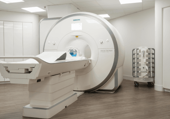
Magnetic Resonance Imaging (MRI): A Closer Look
Functional MRI (fMRI) in Brain Studies
Functional MRI, or fMRI, is a special type of MRI that measures and maps the brain's activity. It helps doctors understand which parts of the brain are involved in different tasks. This technique is especially useful in research and in planning surgeries for brain disorders.
MRI in Oncology
MRI is a powerful tool in the fight against cancer. It provides detailed images of soft tissues, making it easier to detect tumors and monitor their growth. Doctors use MRI to plan treatments and check how well therapies are working.
Advancements in MRI Technology
Recent advancements in MRI technology have made scans faster and more accurate. New techniques allow for clearer images and can even show how tissues are functioning. These improvements help doctors make better diagnoses and treatment plans.
MRI is a key tool in modern medicine, offering detailed images without the use of radiation. Its ability to provide high-resolution images of the body's internal structures makes it invaluable in diagnosing and treating various conditions.
Computed Tomography (CT) Scans: Precision and Speed
CT Angiography for Cardiovascular Health
CT Angiography (CTA) is a noninvasive method to visualize the blood vessels. It uses a special dye and advanced CT technology to capture detailed images of the heart and blood vessels. This technique helps in detecting blockages, aneurysms, and other cardiovascular conditions early, often before symptoms appear.
Low-Dose CT for Lung Cancer Screening
Low-Dose CT (LDCT) scans are used for early detection of lung cancer, especially in high-risk individuals like heavy smokers. This method uses lower amounts of radiation compared to traditional CT scans, making it safer for regular screening. LDCT can identify small nodules or abnormalities in the lungs that might be missed by other imaging techniques.
Technological Innovations in CT Imaging
Recent advancements in CT technology have significantly improved the speed and accuracy of scans. Modern CT scanners can produce high-resolution images in just a few minutes, reducing the time patients need to stay still. These innovations also include better image processing software and lower radiation doses, enhancing patient safety and diagnostic precision.
CT scans are a cornerstone in modern medical diagnostics, offering quick and detailed insights into various health conditions.
Positron Emission Tomography (PET) Scans: Unveiling Metabolic Processes
PET in Neurology
A PET scan is a non-invasive imaging test that helps visualize the metabolic processes in the body. Using a small amount of radioactive material, usually injected into a vein, combined with glucose, the scan captures images of the distribution of this mix as it’s consumed by cells in the body. Areas with higher metabolic activity will take up more of this material — a pattern that can signify various conditions.
PET-CT in Oncology
Cancer cells often have a higher metabolic rate than normal cells. A PET scan can highlight these areas, even when they are too small to be seen by other imaging methods. This early detection can significantly increase the chances of successful treatment and full recovery.
Future Directions for PET Imaging
Medical technology has progressed tremendously over the decades, giving clinicians advanced tools to detect and diagnose various conditions. Among these tools, the PET (Positron Emission Tomography) scan stands out as one of the most powerful diagnostic imaging techniques. Its ability to provide comprehensive insights into the body’s internal functioning is revolutionizing the way medical professionals approach numerous health concerns.
Emerging Imaging Techniques in Cardiovascular Diagnostics
Intravascular Ultrasound (IVUS)
Intravascular Ultrasound (IVUS) is a cutting-edge technique that provides detailed images of the inside of blood vessels. This method uses a tiny ultrasound probe attached to a catheter, which is then inserted into the blood vessels. IVUS is particularly useful for assessing the buildup of plaque in arteries and can help guide treatments like stenting or angioplasty.
Optical Coherence Tomography (OCT)
Optical Coherence Tomography (OCT) offers high-resolution images of blood vessels by using light waves. This technique is similar to IVUS but provides even finer details, making it easier to spot small plaques or other abnormalities. OCT is often used in combination with other imaging methods to give a comprehensive view of cardiovascular health.
Cardiac Magnetic Resonance (CMR)
Cardiac Magnetic Resonance (CMR) imaging is a non-invasive method that uses magnetic fields and radio waves to create detailed images of the heart. CMR is excellent for evaluating heart structure and function, and it can also measure blood flow and detect scar tissue. This technique is invaluable for diagnosing a range of heart conditions, from congenital heart disease to cardiomyopathies.
Emerging imaging techniques like IVUS, OCT, and CMR are revolutionizing cardiovascular diagnostics, offering more precise and detailed images than ever before. These advancements are crucial for early detection and treatment of heart diseases.
Conclusion
In conclusion, advanced imaging techniques have become a cornerstone in modern medical diagnostics. From ultrasonography to PET scans, these technologies allow doctors to see inside the human body with incredible detail. This helps in diagnosing and treating various health conditions more accurately and efficiently. As technology continues to evolve, we can expect even more innovative tools that will further improve patient care and outcomes. The future of medical diagnostics looks promising, with advanced imaging leading the way.
Frequently Asked Questions
What is medical imaging?
Medical imaging is a way to look inside the body using different technologies like X-rays, MRIs, CT scans, and ultrasounds. It helps doctors see what's going on inside without needing surgery.
How has medical imaging changed over time?
Medical imaging has come a long way. It started with simple X-rays and now includes advanced techniques like 3D imaging and MRIs that can show detailed pictures of the body's inside.
What is an MRI used for?
An MRI, or Magnetic Resonance Imaging, is used to take detailed pictures of organs and tissues inside the body. It's often used to look at the brain, spine, and joints.
How does a CT scan work?
A CT scan uses X-rays and a computer to create detailed images of the inside of the body. It's very fast and can show a lot of detail, making it useful for diagnosing many conditions.
What is a PET scan?
A PET scan, or Positron Emission Tomography scan, shows how organs and tissues are working. It's often used to detect cancer and see how far it has spread.
Are there new technologies in medical imaging?
Yes, new technologies are always being developed. Some of the latest include 3D and 4D imaging, which provide even more detailed pictures, and new types of scans that can show how the body is working in real-time.